Young Investigators
Substanzielle Förderung für junge Talente
Mit dem Young Investigators-Programm fördert die Universität des Saarlandes exzellente Nachwuchswissenschaftlerinnen und Nachwuchswissenschaftler aus dem Schwerpunkt BioMed.
Um sich voll und ganz auf Ihre Forschung konzentrieren zu können, erhalten die Geförderten drei Jahre lang finanzielle Unterstützung für ihre Vorhaben. Zudem schafft die Anschubförderung ihnen den nötigen Freiraum, weitere Drittmittel einzuwerben.
Bislang haben fünfzehn Forscherinnen und Forscher aus Saarbrücken und Homburg, die in einem wissenschaftsgeleiteten Wettbewerb ausgewählt wurden, eine Förderung im Rahmen des Young Investigators-Programm erhalten.
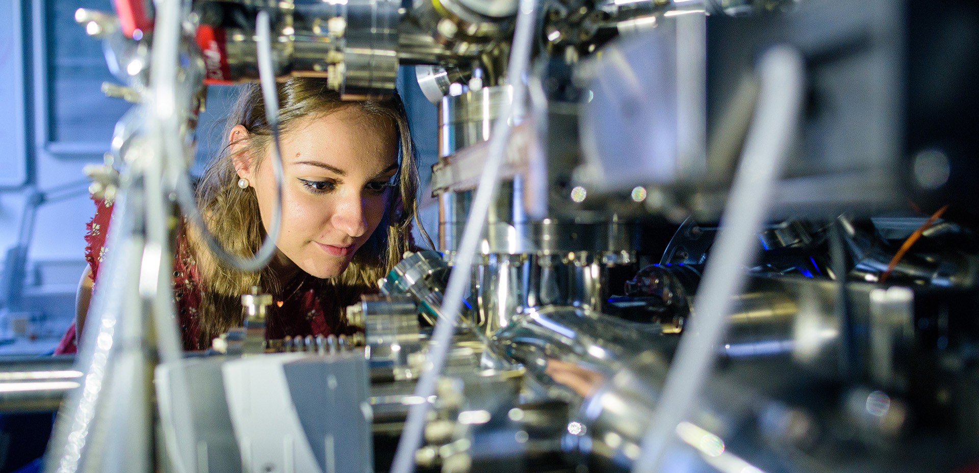
Lattice Dynamics in Microtubule Bundles
Jun.-Prof. Dr. Laura Aradilla Zapata
Microtubules are cytoskeletal filaments spanning the intracellular space of most cells. They can be organized into highly diverse network architectures, reflecting their manifold functions. One of their most striking characteristics is their ability to dynamically grow and shrink by addition or loss of subunits at the tips. In contrast to microtubule tips, the lattice far from the tips was long considered to be static.
Recent findings challenge this paradigm: I could show that the microtubule lattice is dynamic and constantly renews itself by continuously losing and incorporating subunits from the surrounding solution. This surprising property has profound effects on the overall microtubule behavior and characteristics: It influences microtubule tip dynamics, lifetime, resistance to mechanical stress and the activity and localization of other proteins. Yet, many details of lattice dynamics remain elusive. In particular, the interplay between lattice dynamics, mechanics and different microtubule network architectures is largely unknown.
As the shape and functions of individual cells and entire organs depend on the organization of the microtubule cytoskeleton, an improved understanding of the structural and functional aspects of microtubule lattice dynamics depending on network architecture would contribute to a refined comprehension of many physiological and pathological processes associated with the microtubule cytoskeleton.
Here, I propose the investigation of lattice dynamics in microtubule bundles, a highly organized microtubule architecture present in diverse cellular contexts. We will explore lattice dynamics in bundles mediated by crosslinker proteins in three complementary assays:
- In in vitro microtubule bundles,
- in mechanically stressed bundles and
- in neuronal axons, where microtubule bundles represent the structural backbone of intracellular transport and ensure axon consolidation and maintenance.
Overall, this project further strengthens the CRC 1027, fosters collaborations both within and outside of Saarland University and opens up new perspectives for future collaborative research initiatives. Furthermore, it encompasses the investigation of phenomena at interfaces at the cellular and subcellular level and is therefore well aligned with the scope of the upcoming Cluster of Excellence: The organization and mechanics of the cytoskeleton can be considered phenomena of the interface between this highly versatile network and the intra- as well as extracellular environment. In addition, the project links molecular scale phenomena to cytoskeletal organization, mechanics and dynamics and is thus based at the interface between different scales – from molecules to cells.
Weitere Projekte im Young Investigators-Programm

A highly homeostatic extracellular microenvironment is prerequisite for the proper neural activity in the central nervous system (CNS), which is ensured by the blood-brain barrier (BBB), the biointerface between the brain and the peripheral circulation. The BBB is a multicellular structure composed of endothelial cells (ECs), pericytes, and astrocytes. In addition, these cells provide the molecular substrate for migratory oligodendrocyte precursor cells (OPCs) along the vasculature during development. Failure in the recruitment of any of these cell types leads to BBB dysfunction. For instance, abnormal clustering of OPCs on the BV induces BBB breakdown, due to the contact loss of the astrocyte endfeet on the BV.
The OPC migration is mediated by cytokines as well as neurotransmitters. In vitro, using an OPC cell line, it has been shown that GABA activates GABABRs to stimulate migration. In addition, GABA derived from ECs directs interneuron migration as well, and also blocking GABA release impairs BBB integrity. Therefore, we hypothesized that EC-derived GABA guides OPC migration through GABABR signaling and such interaction promotes BBB barrier function.
To substantiate this hypothesis, we investigated OPC migration in the OPC-specific GABABR conditional knockout mice (cKO). Our preliminary data showed that in the dorsal cortex of cKO mice, the developmental expansion of OPCs at the first postnatal week was retarded while the density of these cells on the BV was increased, suggesting decelerated migration and impeded dissociation of OPCs from the BV. Furthermore, we also observed BBB leakage and sustained neuroinflammation in the cKO mouse cortex from the early postnatal week till adulthood, highly likely due to the loss of astrocyte endfeet support on the ECs. Concomitantly, a dysregulated cortical neural circuit was observed in these mice exhibiting increased excitation while suppressed inhibition.
Hence, in this proposed project, we will address the questions whether and how OPCs communicate with ECs via GABABRs during migration and how such interaction influences BBB function. For that purpose, we will investigate the GABAergic communication between ECs and OPCs employing ex vivo slice imaging. We will assess how migratory OPCs interact with ECs and astrocyte endfeet via GABABRs, using two-photon imaging in the living mouse. Finally, we will decipher the intracellular signaling pathways of GABABRs involved in the OPC-EC interaction and the BBB integrity. BBB establishment occurs during the development as well as in the repairing phase of many CNS diseases. Therefore, our study will not only bring novel insights to delineate the mechanism of BBB integrity but also guide a potential strategy for the clinical treatments against CNS diseases.

The central nervous system (CNS) plays a central role to control our body function. The CNS is normally protected against any invasions in order to avoid cell damage in the brain and in the spinal cord. However, in case of autoimmune encephalitis diseases such as multiple sclerosis (MS), autoreactive T cells infiltrate into the brain and attack myelin sheet of neurons, resulting in CNS inflammation, demyelination and neurodegeneration. The model for this disease in rodent is the experimental autoimmune encephalomyelitis (EAE), which leads to the paralysis of the animals. Interestingly, the CNS of mice and rats recover after an EAE whereas in human MS follows a chronic neurodegenerative course. This recovery from EAE in rodents rises the attention of the potential neuroprotective regulation.
Previously, we have shown the encephalitogenic T cells, rather than infiltrating the white matter, penetrate the grey matter in which they closely interact with neurons. Intriguingly, this massive T cell infiltration in the CNS is being cleaned up after animals have recovered. We therefore hypothesize that a neuroprotective mechanism takes place under EAE. Our preliminary data shows that entire cell/fragments from apoptotic encephalitogenic T cells are localized in neuronal somata in the pathological brain, indicating that neurons play a role in removing the inflammatory cells. Furthermore, we observed that during the inflammation T cell mitochondria are taken up by neurons. The neuronal mitochondria transfer was demonstrated to be part of neuroprotective mechanism by another study.
We now aim at understanding how autoreactive T cells interact with cortical neurons under inflamed conditions and decipher this interaction down to the molecular level. We will investigate the potential neuroprotective mechanism, in particular analyzing the cause of encephalitogenic T cell death and determine energy restoration in neurons through external mitochondria uptake. Finally, we will evaluate the neuroprotective outcome. We will address these aims by applying state of the art live imaging techniques combined with quantitative analysis of targeted protein and gene expression. We will observe this dynamic interaction and decipher the molecular mechanism of the potential mitochondria transfer pathway at the single cell level and in pathological brain slices. Understanding the interaction between T cells and neurons will reveal molecular cues that offer neuroprotection during autoimmune CNS diseases. We are convinced that our work will contribute to development of new strategies for the treatment of a broad spectrum of autoimmune diseases.
Natural products represent powerful tools searching for novel anti-cancer drugs. Pamamycins belong to the macrodiolide group of polyketide compounds, heterologously expressed in Streptomyces albus J1074 by the Luzhetskyy group. Driven by their potent bioactivities, the proposed study involves a comprehensive biological screening of pamamycins, elucidating their effects on different hallmarks of cancer in tumor cells and macrophages as crucial players of the tumor microenvironment.
Cancer cells modify their microenvironment according to their needs and thus the microenvironment has been shown to strongly influence the efficiency of therapeutic interventions. Therefore, this phenotypic screening aims to identify novel anti-tumor targets and strategies beyond the exclusive killing of malignant cells.
2D and 3D cell culturing models will be employed to assess the in vitro activity profile on tumor cells and macrophages. The most prominent compound candidates will be studied further in co-culturing models and in zebrafish embryos as an emerging in vivo tumor model organism.
In addition, this study aims to focus specifically on the deregulation of cellular energetics as a hallmark of cancer. This includes a profound analysis of pamamycins-induced effects on the metabolism of tumor cells and macrophages. Moreover, metabolic modulation by natural compounds and already known drugs will be studied as an anti-cancer strategy in the context of cancer – macrophage interactions.
The objective of this research project is to investigate how modifications of cell membranes (or the underlying cytoskeleton) modify their surface interactions with the endothelium of blood vessel (membrane-membrane interactions), and influence the in vivo blood flow, with a particular attention to white blood cells (WBC). It has been shown that interaction between WBC and the endothelium are governing their main locomotion, since they are prone to attach to the endothelium and roll on it. Moreover, WBC adhering to the endothelium have a significant effect on the hemodynamics of the vessel.
In order to achieve this objective, I will work with mesentery and dorsal skinfold chamber mouse models. I will both use WBC with artificially modified cytoskeleton and cells from mice with a mutation inducing Chorea-acanthocytosis. This disease is characterized by a mutation of the chorein (VSP13A) which is a structural protein of the cytoskeleton and thus alters the mechanical properties of the cells. In this pathological case, both red blood cells (RBC) and WBC present a higher effective rigidity.
In human subjects, chorea-acanthocytosis is characterized by neurological disorders, such as chorea, parkinsonism or dystonia. It is currently unclear if or how the modification of blood cell membrane properties leads to the apparition of such neurologic disorders. Studying how the membrane modifications alter the cells flow in the circulation would bring an improved understanding of the molecular and cellular disease mechanisms. Moreover, this research will generate fundamental knowledge about the relation of cell membrane properties and blood flow in capillary networks.
In the recent years, we developed and optimized a 3D microfluidic platform that enables fabrication of lipid bilayer that is free‐standing, highly reproducible, horizontal, stable, fluid, solvent‐free, and a physiologically relevant lipidic composition. This superior platform allows the reconstitution of proteins, such as transmembrane, peripheral, and pore‐forming proteins, that can be added to the bilayer in controlled orientation and keep their native mobility and activity. Combining simultaneous optical and electrophysiological methods, we used this platform to determine the nascent pore expansion rate of individual SNARE mediated vesicle fusion with a single SNAREpins resolution.
We aim in this proposal, to use this superior platform to the field of viral fusion. Viral fusion is in many aspects very similar to SNAREs mediated fusion, with the difference that the fusion is mediated by viral proteins The characterization of a viral fusion dynamic pathway can be measured using the same methods that we employ to study SNARE mediated single vesicle fusion. We aim to employ Virus-Like Particles (VLPs) composed of a vesicle containing viral proteins. These VLPs are non-infectious because they contain no viral genetic material.
As a first proof of concept, we aim to characterize and to understand the fusion of Influenza VLPs with our free-standing bilayer. We aim to characterize the nature of the pore and the corresponding pore expansion rates, before to compare these results with stalk free-energy calculations from Prof. J. Hub (UdS). Once, we have demonstrated that our superior platform is suitable to study viral fusion. We aim to study and to understand the fusion dynamic pathway of SARS-CoV-2 VLPs. This is a different fusion process than the case of Influenza, as we need to insert a protein receptor ACE2 into our free-standing bilayer to achieve the fusion process. We aim to understand the biochemical conditions that allow the spontaneous fusion of SARS-CoV-2 VLPs with our free-standing bilayer (enriched with ACE2 receptors). Then, we aim to characterize the nature of the pore and the corresponding pore expansion rates, before to compare these results with stalk free-energy calculations from Prof. J. Hub. If times allow, we aim to measure the fusion rate for different mutations of SARS-CoV-2 spike proteins and extract the effect of these mutations on the fusion energy landscape. Again, these results aim to be compared with results from stalk free-energy calculations of Prof. J. Hub.

The development of new antibiotic agents is a central aspect of infection research and the rise of antibiotic resistances makes this an increasingly urgent matter. Ribosomally synthesized and post-translationally modified peptides (RiPPs) are an emerging superfamily of natural products that often exhibit promising antimicrobial activities and recent studies have demonstrated how RiPPs produced by human commensals can have host protective properties. Indeed, the rise of genome mining for the identification of novel RiPP biosynthetic gene clusters confirms that the potential for producing RiPPs is widespread amongst both beneficial human commensals as well as amongst human pathogens. Thus, it seems promising to take a closer look at the RiPPs present in the human microbiome not only in the search for novel drug leads in the fight against infectious diseases, but also for understanding their roles in and how they affect microbial communities.
The project will therefore initially focus on lead molecules able to overcome antibiotic resistances with the ultimate goal of the development of new anti-infective agents that can be translated into novel treatments. At later stages, the project will be extended to also mine metagenomic datasets to identify new RiPPs from the human microbiome and enable the study of their native functions. Thus, this project exhibits a strong synergy with project IMAGINE of the HIPS-UdS TANDEM initiative, which will provide microbiome and metagenomic datasets from clinical samples that can in turn be prospected towards identifying new, unique natural products.
The initial lead molecules investigated will be the hogocidins. These are class II lanthipeptides with a selective anti-MRSA activity and they originated from a beneficial human commensal isolated from the skin microbiota of a healthy individual. Through hogocidin secretion, the producing strain exhibits microbiome protective features and helps preventing Staphylococcus aureus colonization of the skin. Systematic structure-activity-relationship (SAR) studies will aid in lead structure optimization and will be complemented by genome mining efforts aimed at isolating closely related, naturally occurring homologs in search for superior lead structures and potential probiotics. These efforts will further establish general methodologies and work procedures that will later be utilized for heterologously producing new RiPP-based lead structures identified in the human microbiome.
The project will combine methods from biochemistry, microbiology, chemical biology, medicinal chemistry, analytical chemistry, and bioinformatics.

Research on an interaction between proteins and cell membranes is still veiled, although it is the most basic mechanism in membrane-mediated cellular processes, including antimicrobial peptide action and peptide-mediated cell membrane penetration. Recently, we studied protein-like dynamic polymers (biodynamer) of hexaethylene glycol conjugated carbazole dialdehydes, and amino acid hydrazides.
We demonstrated their strong interactions with cell membranes similar to functional peptides like cellpenetrating peptides (CPPs) or antimicrobial peptides (AMPs). Also, their superior tunability controlling their characteristics makes them a suitable research tool for investigating peptide/membrane interaction.
In this project, we aim to develop oligomeric biodynamer, oligodynamer to study various physicochemical factors affecting cell-membrane interactions, and design a new class of functional peptides more easily controllable than conventional peptides. For this purpose, we sophisticatedly control physicochemical properties of oligodynamers, such as structure, rigidity, charge, and hydrophobicity, which are expected to affect the interaction. We will investigate their effect on the oligomer/membrane interaction using microscopies, calorimetry, and the Förster resonance energy transfer (FRET) technique. We expect this project expands the applicability and potential of dynamic polymer series in biomedical fields.

For decades, neutrophils were considered as a homogeneous population of short-lived circulating and tissue patrolling immune cells that possess antimicrobial functions. However, with the emerging evidence of divers neutrophil sub-populations in health and disease with functionally different phenotypes, this long-believed traditional dogma has outdated. Surface markers, density, maturity, morphology, functional aspects and localization characterize the heterogeneity of neutrophils. However, little is known about the contribution of neutrophil subsets during viral infection-related carcinogenesis although infections with high-risk human papillomaviruses (HPV) are on the rise to the major etiologic agent for cancer at mucosal bio-interfaces.
Oncoproteins encoded by HPV have the potential to critically modulate the landscape of neutrophil plasticity and the efficacy of immunotherapeutic approaches. Notably, neutrophils are crucial for the efficacy of antibody-based immunotherapeutic approaches and thus likely affected by subset transition towards "unfavorable" neutrophils. IgA antibodies effectively engage neutrophils for antibody-dependent cellmediated cytotoxicity (ADCC) of tumor cells. We recently developed an impedance-based assay to test neutrophil activation for ADCC of adherent target cells. This assay allows an uninterrupted longterm real-time monitoring of kinetics accompanied by the detection of critical mediator release. Changes in kinetics and ADCC efficacy upon modulation of the target cells by HPV-oncoprotein expression or of the effector cells due to priming with a HPV-driven cancer cell instructed tumor microenvironment (TME) can be monitored in detail.
The aim of the suggested project is to analyze how natural occurring, and tumor virus-infection TME-induced neutrophil subsets affect antibodybased immunotherapeutic approaches and how unfavorable alterations could be reverted by novel pharmacological compounds. Compounds capable to revert TME-related neutrophil subset transition could support immunotherapeutic approaches even in a broader perspective for malignant and inflammatory diseases. In summary, investigating how viral infection-related cancer instructed TME affects neutrophil subset transition at a single cell level combined with protein activity and functional read outs will help on the one side to identify promising markers for patient’s risk and therapy stratification and on the other side to identify promising novel small molecules for potential clinical development.
The ocular surface is an immune privileged site that was long thought to be devoid of microbiota in the absence of infection. However, with the onset of next generation sequencing (NGS) as a major method for analysis of microbiomes, there is sound evidence demonstrating the colonization of the ocular surface with commensals in the healthy eye. Moreover, the composition of the eye microbiome has been shown to be influenced by age, drug treatment and diseases. Likewise, the expression of smallRNAs e.g. microRNAs (miRNAs) in the conjunctiva and also the stroma and epithelial cells of the cornea has been shown to be altered in genetic eye conditions as for example congenital aniridia or keratoconus. Treatment of these conditions often require the replacement of the affected cornea by PKP with adjuvant local immunosuppressive therapy that might influence the local microbiota. Moreover, recent evidence suggests that miRNAs also have the potential to regulate gene expression across species and genera, for example by influencing gene expression in certain commensal bacteria, which in turn can have an effect on the host.
The aim of the proposed project is to shed further light onto the host-microbiome interaction via miRNAs using the ocular surface with its limited bacterial diversity as model system. Conjunctival miRNA expression will be analyzed in conjunction with the microbiome in the same individuals in a longitudinal fashion by using smallRNA seq and metagenomic sequencing. Samples will be collected from patients with keratoconus undergoing penetrating keratoplasty over the course of therapy and healthy volunteers over a respective time interval to answer the following questions:
- Is there a correlation between the expression of specific miRNAs and the presence of specific bacterial species in the healthy eye or the diseased eye? and
- How is the miRNA expression and/or the local microbiome affected by penetrating keratoplasty in the course of topical antibiotic and corticosteroid treatment?
In addition, in vitro testing of selected host-microbiome interactions will be performed as proof of concept for the miRNA driven interaction of human cells with the microbiome. This project can provide the foundation for future studies focusing on the host-microbiome interactions at other, more complex sites such as gut or skin.
Most living cells are supported by a complex medium consisting of a network of collagen fibers known as the extracellular matrix (ECM). Its physical parameters, such as the network density and the mechanical stiffness, can differ from one tissue to another and impacts the functions of the cells that it contains, such as their migration behaviour. Cells migrate through dense ECMs by creating channels, whereas if the density is low, they leave the ECM intact and squeeze through holes and pores of the network. This mechanism is experimentally documented cells create protrusion which attach to the fibers of the network. Recent numerical models are able to accurately describe this mechanism.
Over the past decades, many experiments have shown that some cells can deform and stiffen the surrounding ECM by contraction. Such deformations are used as a means of communication between cells as some are sensitive to the mechanical stiffening of the ECM. Collagen-mediated forces between cells can be transmitted over distances ranging from a few micrometers to a few hundreds of micrometers.
To understand the mechanics of long-range deformation in fibrillar networks, several models have been investigated and are supported by experimental studies. Recently however, Pakshir et al. have experimentally shown that macrophages may not only sense deformation gradients, but also the rate of the deformation. The sensitivity to the dynamics of the matrix (like temporal changes in the local strain rate or even low-frequency vibrations) seems to enable even more remote cells to be guided towards the contractile cell, which is a very new experimental observation. No proper theoretical and numerical study of this phenomenon has been performed so far, which is what we are aiming at in this project.

Epigenetic signatures regulate how the genetic code inscribed in DNA sequence is interpreted and thus govern the state and function of each individual cell, ultimately giving rise to hundreds of different, highly specialized cell types. Understanding gene regulation is therefore paramount for understanding normal cell development as well as aberrant cellular phenotypes in disease.
In this project, we will develop a framework of computational methods to define epigenetic cell states associated with changes in the cell environment and to dissect the regulatory dynamics involved in the immune response to pathogen exposure. Our collaboration partners are generating single-cell, multiomics profiles of human immune cell states in naïve samples and samples challenged with exposure to different pathogens, including HIV, flu, MRSA/MSSA, Anthrax and Organophosphates.
We will develop and apply methods for the integrative analysis of epigenomic data by modeling the interplay between DNA sequence, DNA methylation, chromatin accessibility and gene expression. We will identify and characterize genomic elements involved in the epigenetic regulation of cell state and we will derive celltype-specific, multi-omics biomarkers of pathogen exposure. Integrating DNA sequence, epigenetic profiles and regulator expression, we will quantify and compare the activity of transcription factors (TFs). The methods developed in this project will be made available in user-friendly and scalable software tools.
The project directly synergizes with ongoing efforts at UdS in the NanoBioMed area that emphasize single-cell approaches and contribute to enhanced understanding of gene regulation. Our computational methods are directly transferable to other systems and tissues involving dynamic regulation of cell state, including changes induced by metabolism, drug-based perturbations, microbiome and cellular interfaces
Short-term plasticity of synaptic transmission is a key step of information processing in the brain. Ca2+ plays a prominent role in many use-dependent forms of plasticity. Recently, we could assign a crucial function to non-selective, Ca2+-permeable TRPC5 channels in the control of presynaptic Ca2+-dynam-ics and synaptic facilitation of primary hippocampal neurons. Specifically, we could show that the combined loss of TRPC1/C4/C5 channels reduces the action potential evoked response, the readily releasable pool size and speeds up synaptic depression on response to high frequency stimulation (HFS). Expression of TRPC channels turned classic synaptic depression into short-term enhancement of synaptic signaling. Furthermore, TRPC channels are able to prolong the presynaptic Ca2+-rise in response to trains of action potentials, showing for the first time that TRPC channels provide an additional presynaptic Ca2+ entry pathway.
Importantly, hippocampal neurons expressing endogenous TRPC5 channels showed strong short-term enhancement of synaptic signaling during HFS. Collec-tively, these results identify TRPC channels as key mediators of presynaptic Ca2+-homeostasis capable of regulating synaptic strength and plasticity. Preliminary studies using the new TRPC5-GFP knock-in (ki) mouse line (in collab. U. Böhm, UdS) show that granule cells of the area dentate express TRPC5. Mossy fiber synapses of granule cells with pyramidal cells of the CA3 region show a pronounced short-term facilitation, the molecular mechanisms of which are only poorly understood. In the proposed project, optogenetic Ca2+-indicators, as well as electrophysiological measurements will be used to investigate the possible influence of TRPC channels on the plasticity behavior of mossy fiber synapses in acute brain slice preparations. Together with the research groups Rettig, Kirchhoff, Flockerzi and Böhm, the synaptic transmission of Mossy fiber synapses from TRPC-deficient and TRPC4-GFP ki mouse lines will comparatively analyzed. These experiments promise new insights into central mechanisms of synaptic short-term plasticity in the intact neuron-glia network.

Understanding the physical processes at the membrane of cells is of great scientific and technological importance. Examples include transport of sub-nanometer particles across the membrane channels, conformational deformations of the cell membrane to internalize larger particles, and formation of microtubule- or actin-based protrusions.
In this proposed project, we aim at understanding the mechanisms of two distinct processes:
- formation and growth of microtentacles (McTNs) and
- wrapping of nanoparticles by lipid membranes.
McTNs are one-dimensional microtubule-based protrusions that are generated by circulating tumor cells (CTCs) and contribute to cell adhesion and aggregation. CTCs play a crucial role in the spread of tumors during metastasis. There are anti-cancer drugs that stabilize microtubules or depolymerize f-actin to block the progression of mitosis and prevent cell division of cancerous tumor cells. However, the main drawback of this type of drugs is that they promote the formation of McTNs (by weakening the actin cortex against growing microtubules), thus, increase the chance of metastasis.
In a preliminary study in collaboration with the groups of Profs. Lautenschläger and Santen, we have managed to generate McTNs in non-cancerous cells using specific drugs that influence the actin cortex. We will further develop and automate our image processing package for 3D reconstruction of cells, which enables us to systematically characterize the structure and dynamics of McTNs for various types and concentrations of drugs. We will also develop a model for microtubule bundle growth and stabilization against barriers and study the mechanisms of McTN formation under the influence of drugs. The long-term aim is to better understand the conditions initiating metastasis and to design more efficient drugs and strategies for cancer treatment.
Our second major goal is to elucidate the mechanism of wrapping of nanoparticles by lipid membranes. In a related study, we previously clarified the role of endo/exocytosis rates and microtubule-assisted transport on receptor clustering at the mammalian cell surface with the group of Prof. Schmitt. Here, we plan to solely focus on phagocytosis and numerically investigate how the interplay between particle properties (such as its size, shape, and softness), elasticity of the membrane and actin cortex, particle-membrane adhesion, and coupling between actin cortex and plasma membrane determines the success rate of wrapping. We also expect that the contribution of actin polymerization at the leading edge of the membrane is influential. The simulation results will be validated by comparing them to two types of experiments: imaging single phagocytosis events at the cell membrane, or statistically evaluating the internalization efficiency of cells upon varying the key parameters in an environment containing many target particles. Phagocytosis is of vital importance for fighting infection by neutrophils. Thus, in a broader perspective, investigation of their wrapping mechanism is in line with other extensive studies at Saarland University on the mechanisms of search, response, and clearance in the immune system. The results may also help to design novel drug delivery strategies.
Our proposed study potentially suggests new experiments on cell membranes and can trigger new collaborations with other research groups of the university.
This proposal aims to investigate adaptive immune responses at epithelial interfaces in the context of infections, autoimmune diseases and cancers at the single cell level.
Immune responses will be characterized, the influence of proinflammatory modulators like Progranulin-antibodies will be studied on responses against infections e.g. COVID19. Possible contribution of infectious agents to cancerous transformation will be studied. Novel targets at epithelial interfaces will be identified by proteomic approaches such as modified SEREX, screening of (posttranslationally) protein arrays, SERPA with autologous patient sera and recombinant BCRs, characterized and subsequently used for immunotherapeutic approaches in cancer, infectious diseases and autoimmune diseases. These BCRcomponents can be incorporated into IgG antibodies, immunotoxins, bispecific antibodies or CAR-T cells. This innovative technique is independent of the MHC-I machinery. Highly neutralizing, super affine monoclonal antibodies can be used for passive immunization against infectious diseases.
The key for this proposal is the expertise in finding and characterizing these therapeutic targets. These techniques and platforms can be used for a variety of relevant, today still life-threatening diseases, providing the possibility for the basis of multiple new research proposals, contribution to consortia as well as start ups.